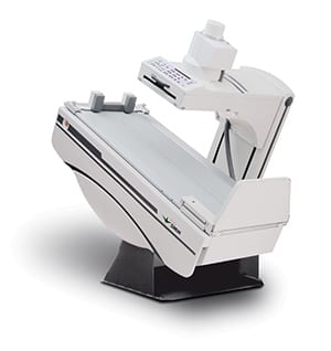
Fluoroscopy is an imaging technique commonly used to obtain real-time images of the internal structures of a patient through the use of a fluoroscope.
For this imaging method , a patient is placed between an x-ray source and a fluorescent screen; an x-ray image intensifier is employed to minimize radiation exposure and a CCD camera allows the images to be played and recorded on a monitor. The result is much like an x-ray "movie”; the x-ray beam is continually passed through the body part being examined, and is transmitted to the monitor so that the body part and its motion can be seen in detail.
In general, fluoroscopy has a broad application in radiologic procedures. It is often used in studies of the gastrointestinal tract, including those involving the use of barium, arthrograms, orthopedic surgery, urological exams and surgery, and angiography of the leg, heart and cerebral vessels. It can also aid in the placement of cardiac rhythm management devices such as pacemakers and implantable cardioverter defibrillators.
A wide variety of catheter placement procedures also involve the use of a fluoroscope. Some preparation may be involved for your exam; you will be given specific instructions at the time of scheduling.

Fully Accredited by the
American College of Radiology


