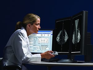
MRI, short for Magnetic Resonance Imaging, combines the use of radio frequencies and a powerful magnetic field to align the hydrogen atoms in your body, which produces an extremely detailed internal image. Images of the soft tissues in your body are often captured through MRI because it produces good contrast. Breast Imaging is routinely done with Mammography or Ultrasound; however, if your doctor needs more information than a mammogram or ultrasound can normally reveal, he or she may perform a Breast MRI.
As a diagnostic tool, Breast MRI is most effective when used in addition to a mammogram or another breast imaging exam.
MRI of the breast is used for the following:
- evaluating abnormalities detected by mammography.
- identifying early breast cancer not detected through other means,
especially in women with dense breast tissue and those at high risk for the disease. - screening for cancer in women who have implants or scar tissue that might jeopardize an accurate result from a mammogram.
- determining the integrity of breast implants.
- distinguishing between scar tissue and recurrent tumors.
- assessing multiple tumor locations.
- looking for multiple tumors prior to breast conservation surgery.
- determining whether cancer detected by mammography or ultrasound has spread further in the breast or into the chest wall.
- determining how much cancer has spread beyond the surgical site after a breast biopsy or lumpectomy.
- assessing the effect of chemotherapy.
- providing additional information on a diseased breast to make treatment decisions.

Fully Accredited by the
American College of Radiology